
Application of High-Frequency Handheld Ultrasound in the Medical Aesthetics Industry (Part II)
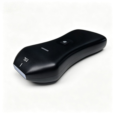
In recent years, the medical aesthetics industry has shifted from extensive and experience-based operations to a refined, visualized and personalized diagnosis and treatment model. Traditional aesthetics medical treatment relies heavily on physicians' visual observation, manual palpation and clinical experience to judge subcutaneous anatomical layers.This model has many inherent limitations: large errors in dermal thickness assessment, low recognition of subcutaneous blood vessels and fascial layers, inability to identify the properties of previously injected fillers, and difficulty in precisely controlling the depth of photoelectric/injection energy therapies.The above problems can easily induce complications such as vascular embolism, facial uneven deformity, nerve damage and unsatisfactory treatment outcomes. In addition the lack of standardized quantitative methods to compare pre-operative and post-operative treatment outcomes has become a core difficulty hindering the safe and standardized implementation of medical aesthetic diagnosis and treatment.
High-frequency medical ultrasound boasts core advantages including non-invasive real-time imaging, zero ionizing radiation, convenient bedside operation, and high resolution for soft tissue imaging. It can clearly delineate delicate soft tissue anatomical structures such as the epidermis, dermis, subcutaneous fat, SMAS fascia, superficial blood vessels and tiny facial nerve branches, filling the subcutaneous blind zones invisible to the naked eye and undetectable by manual palpation — yet the lack of its standardized clinical application has become a core obstacle restricting the safe and standardized delivery of aesthetic medical treatment.
Mainstream high-frequency linear ultrasound probes for clinical use (conventional frequency: 7–18 MHz). Such probes are suitable for superficial soft tissue imaging in medical aesthetics and boast remarkable clinical application value, covering the whole process of preoperative assessment, intraoperative guidance and postoperative review:
Preoperative imaging evaluation
Accurately measure the thickness of each layer including the skin, subcutaneous fat, fascia and muscles; distinguish true puffy eyes from pseudo soft tissue bulging; identify abnormal tissues such as unknown residual materials from previous injections and subcutaneous cystic nodules
Intraoperative real-time guidance
Dynamically display the course of superficial facial arteries and veins to assist physicians in avoiding blood vessels during puncture; track the penetration depth of the needle in real time to fundamentally eliminate the risk of vascular embolism caused by injection materials entering the bloodstream.
Postoperative follow-up monitoring
Screen for delayed subcutaneous hematoma and filler displacement, continuously monitor local inflammatory responses, and detect postoperative complications in a timely manner.
Cheek (the large area of the face on either side)
Common issues in the frontal region: sagging and drooping cheek areas, baby fat or excessive fat deposits
Precautions for medical aesthetics: parotid gland boundary, course of facial blood vessels
Mobile Supermarket Adaptation Project:
-
Baby fat or excessive fat: Preoperative ultrasound is used to expand the operative visual field, distinguish the boundary between superficial subcutaneous fat and parotid fat, identify deep vascular plexuses, and avoid massive hemorrhage and accidental parotid gland injury during liposuction and lipolysis. Postoperative follow-up ultrasound can serve as the primary examination method.
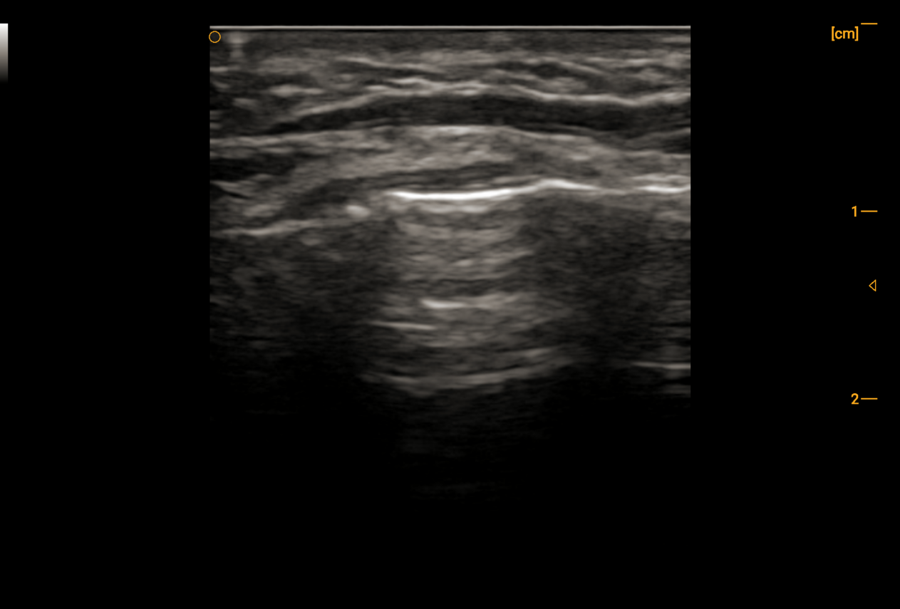
Masseter region (cheek above the mandibular angle)
Common midface concerns: enlarged masseter muscles, broad and square facial contours
Precautions for Medical Aesthetics: Masseter muscle thickness and boundaries, facial arteries and veins, course of facial nerve
Palm Super Adaptation Project:
Masseter hypertrophy: Ultrasound-guided injection of slimming face needle into the planned area, real-time ultrasound monitoring of injection process, and regular ultrasound follow-up of postoperative changes.
-
Square wide face: Osteotomy and bone grinding surgery for enlarged jawbone, with ultrasound monitoring of postoperative changes.
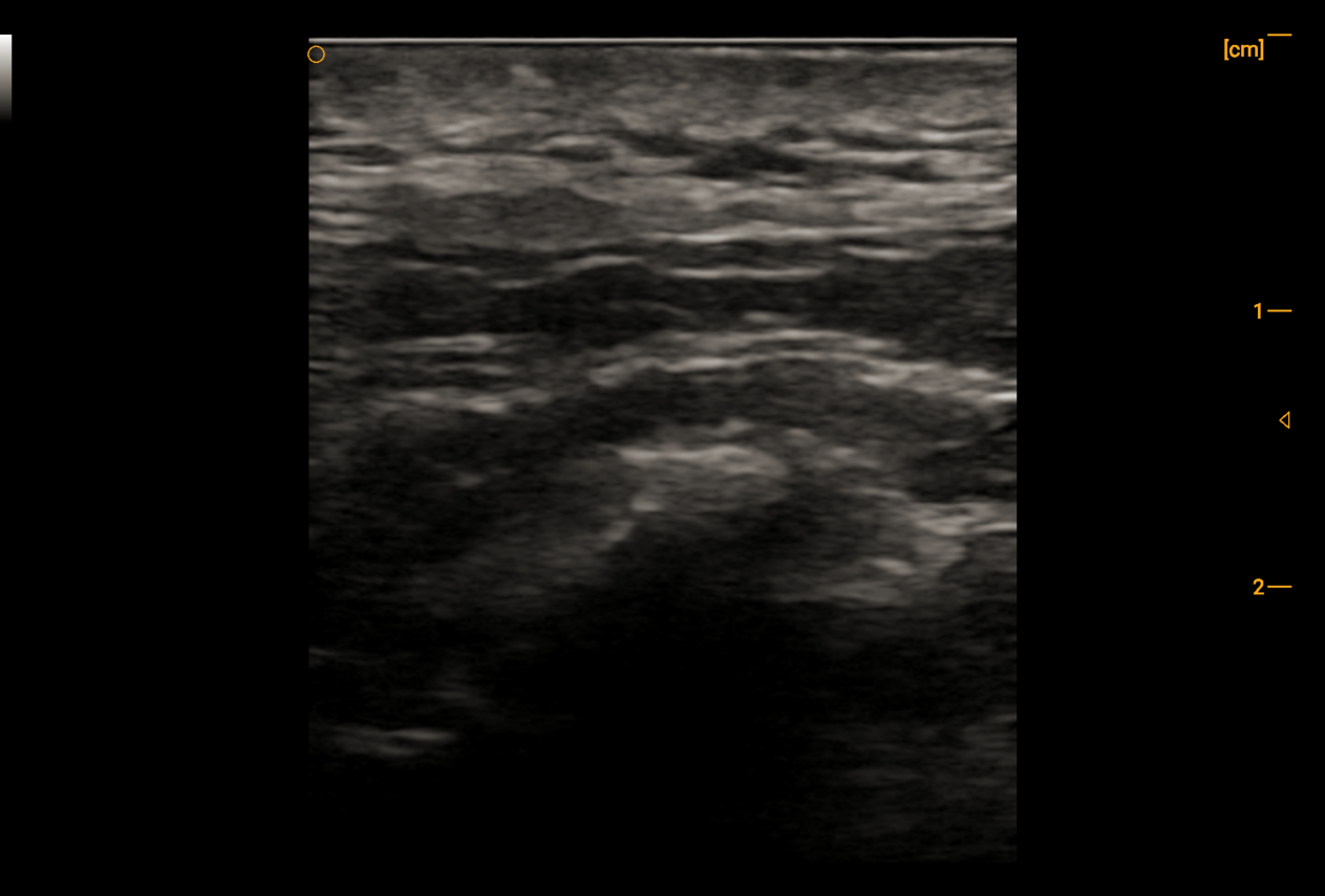
Angular oris region and lips
Common Periorbital Concerns: Elimination of Oral Commissure Lines (Marionette Lines), Lip Contouring
Medical Aesthetic Precautions: Precise Localization of Aberrant Labial Artery Branches and Mental Nerves
Ultrasound Palm Probe Suitable Treatments:
Nasolabial folds and marionette lines: Preoperative ultrasound is used to hierarchically identify four layers including the skin, dermis, orbicularis oris muscle and submucosal glands; it accurately locates aberrant branches of the labial artery, facilitating botulinum toxin injection to relax the depressor anguli oris muscle or hyaluronic acid filling of grooves. Postoperative ultrasound assessment serves as the primary examination modality.
Autologous fat grafting correction for asymmetric lips: Ultrasound measures the bilateral thickness of the orbicularis oris muscle and subcutaneous spaces, precisely calculating the volume difference for grafting, avoiding blood vessels and glands, and reducing postoperative asymmetry and nodule formation.
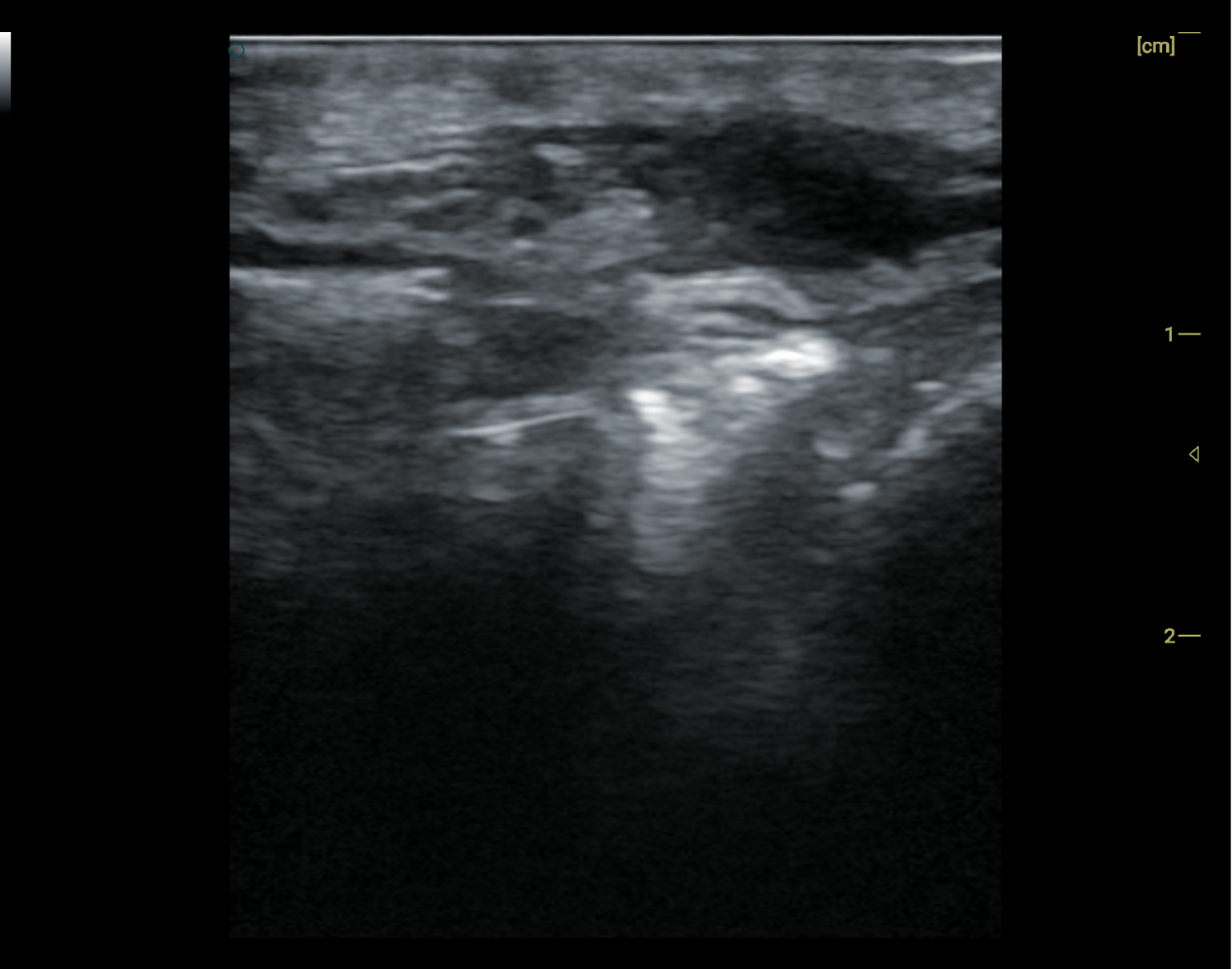
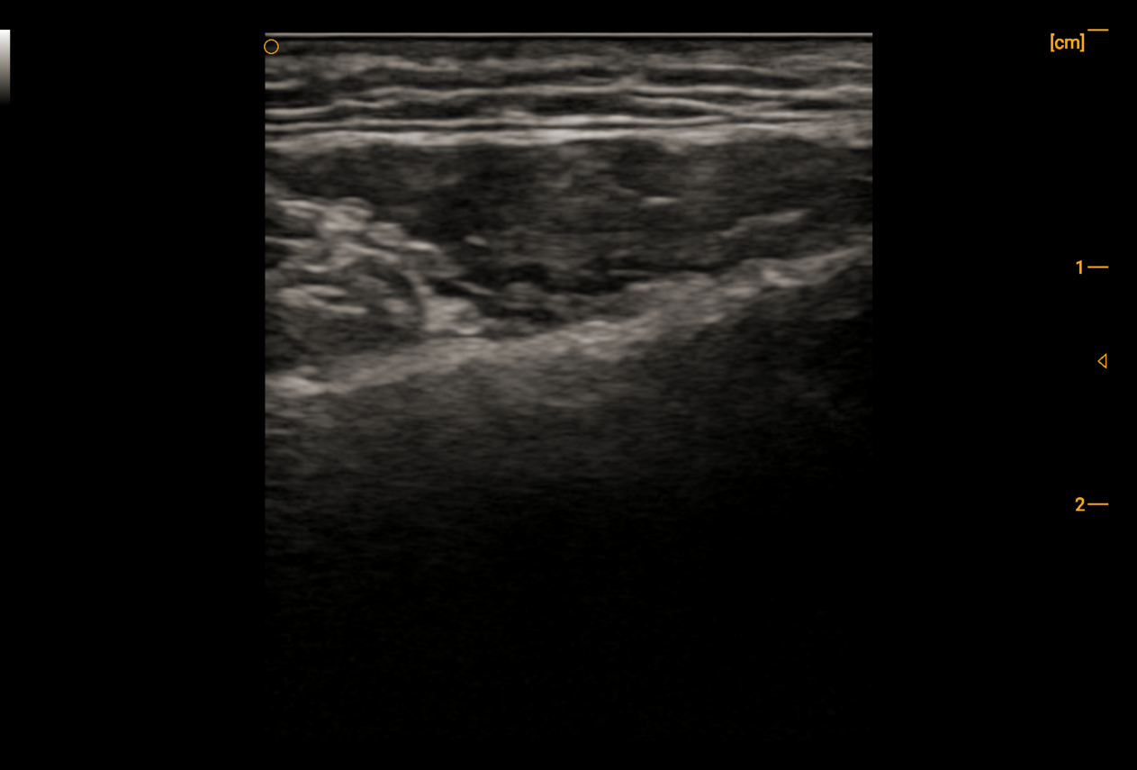
Jawline and Double Chin
Common Nasal Area Concerns: Blurred and Sagging Jawline, Fatty Double Chin, Mixed Sagging and Fat Type
Aesthetic Medical Precautions: Submental Arteries and Veins, Branches of the Facial Nerve, Subcutaneous Fat Thickness
Applicable Palm Ultrasound Treatments:
Blurred and Sagging Jawline: Preoperative ultrasound mapping of the surgical area, real-time intraoperative ultrasound guidance to inject lipolytic agents into the mapped zone with continuous monitoring of injection delivery. Regular postoperative ultrasound follow-ups to assess post-treatment changes.
Double chin (a type of fatness): Preoperative ultrasound is used to map out liposuction or fat dissolving areas, intraoperatively guide liposuction cannulas or fat dissolving injections in real time to avoid vascular plexuses. Ultrasound is the preferred imaging modality for evaluating postoperative changes.
Chin (mental region)
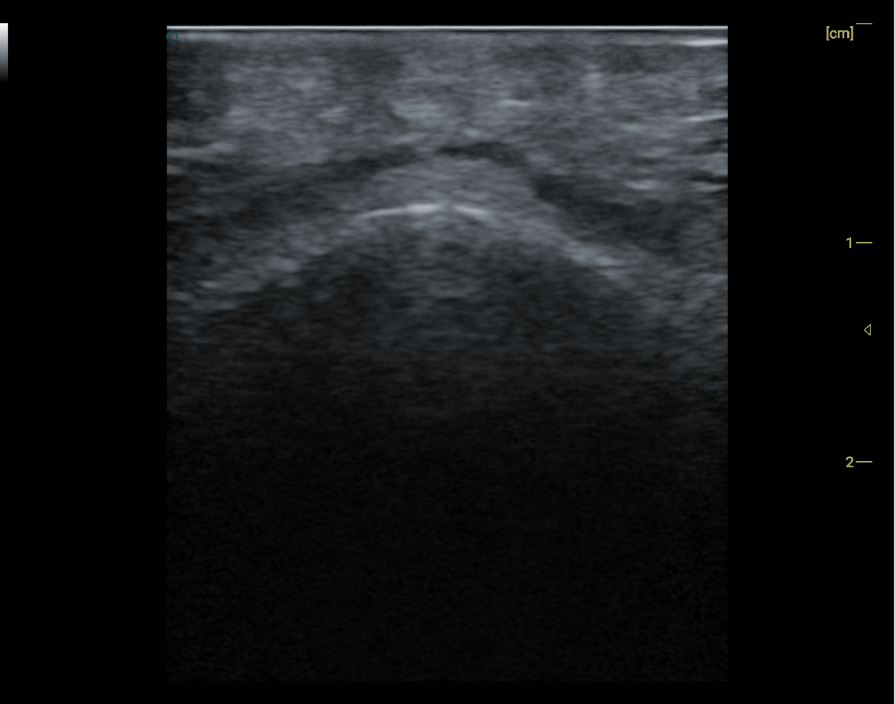
Common Problems: Retruded chin, microgenia, left-right chin asymmetry, compensatory shaping for mild protruding mouth
Medical Aesthetic Precautions: Mental artery and vein, displaced mental foramen with anatomical variation
Applicable Ultrasound Imaging Procedures:
For retruded and short chins: Preoperative ultrasound-based surgical planning, including precisely locating displaced mental foramina with anatomical variations, avoiding blood vessels and nerves, and defining filling boundaries. Real-time monitoring of needle position during surgery to prevent intravascular injection and compression of the mental nerve by fillers; Postoperative screening for delayed hematoma and filler migration causing nerve compression. Regular ultrasound follow-ups to assess filler placement
Temporal region (temple)
Common periorbital concerns: bony temporal depression, prominent cheekbones, soft tissue atrophy, asymmetric temples, lifting of the outer brow, fading of crow's feet
Medical aesthetic precautions: course of superficial temporal artery branches, course of facial nerve
Suitable projects for palm ultrasound:
Wide facial appearance caused by depressions and prominent cheekbones: Precisely locate variant superficial temporal artery branches before surgery and mark safe injection planes. Track the needle path in real time during the procedure to prevent intravascular injection of hyaluronic acid (autologous fat) or penetration into the temporalis muscle. Screen for delayed hematoma and compression of blood vessels and nerves by fillers after surgery.
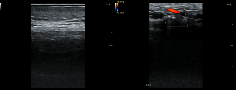
Unlike focused ultrasound therapeutic devices, ultrasound imaging devices take visual imaging as their core function, fully covering the complete diagnosis and treatment cycle of pre-aesthetic surgery assessment, intraoperative navigation and postoperative follow-up. Before surgery, they can quantify thickness parameters of various skin layers, identify different unknown subcutaneous foreign bodies and filler materials, and screen out high-risk anatomical variations of blood vessels in advance. During surgery, they provide real-time guidance for minimally invasive injection, thread lifting implantation, local lipolysis and other procedures, avoiding the risk of vascular and nerve damage from the source. After surgery, they dynamically monitor the whole process of collagen regeneration, subcutaneous edema, delayed hematoma and scar repair, and establish quantifiable and comparable objective imaging evaluation criteria for treatment efficacy. At present, there remain three major pain points restricting the implementation of ultrasound imaging technology in China's medical aesthetic industry: low clinical penetration rate of relevant equipment, insufficient professional image interpretation capabilities of medical aesthetic physicians, and lack of standardized supporting diagnosis and treatment protocols. In view of the above industry status, this paper systematically summarizes the key practical application points of ultrasound imaging in various medical aesthetic scenarios to provide reference for clinical practice.
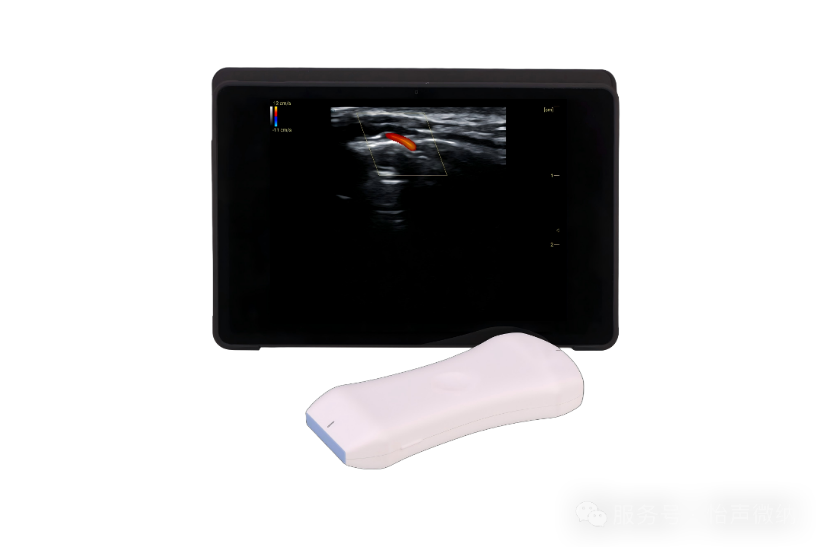
The era of refined visual medical aesthetics has arrived. Ultrasound imaging equipment serves as core facilities for medical institutions to boost competitiveness and reduce complications. Our company offers customized probes and collaborative clinical supporting solutions. We sincerely invite medical device distributors and OEM partners nationwide to discuss cooperation. You may leave a message in the background to obtain detailed cooperation materials, and we look forward to jointly exploring new industry development with you!

